
FIGURE 3.
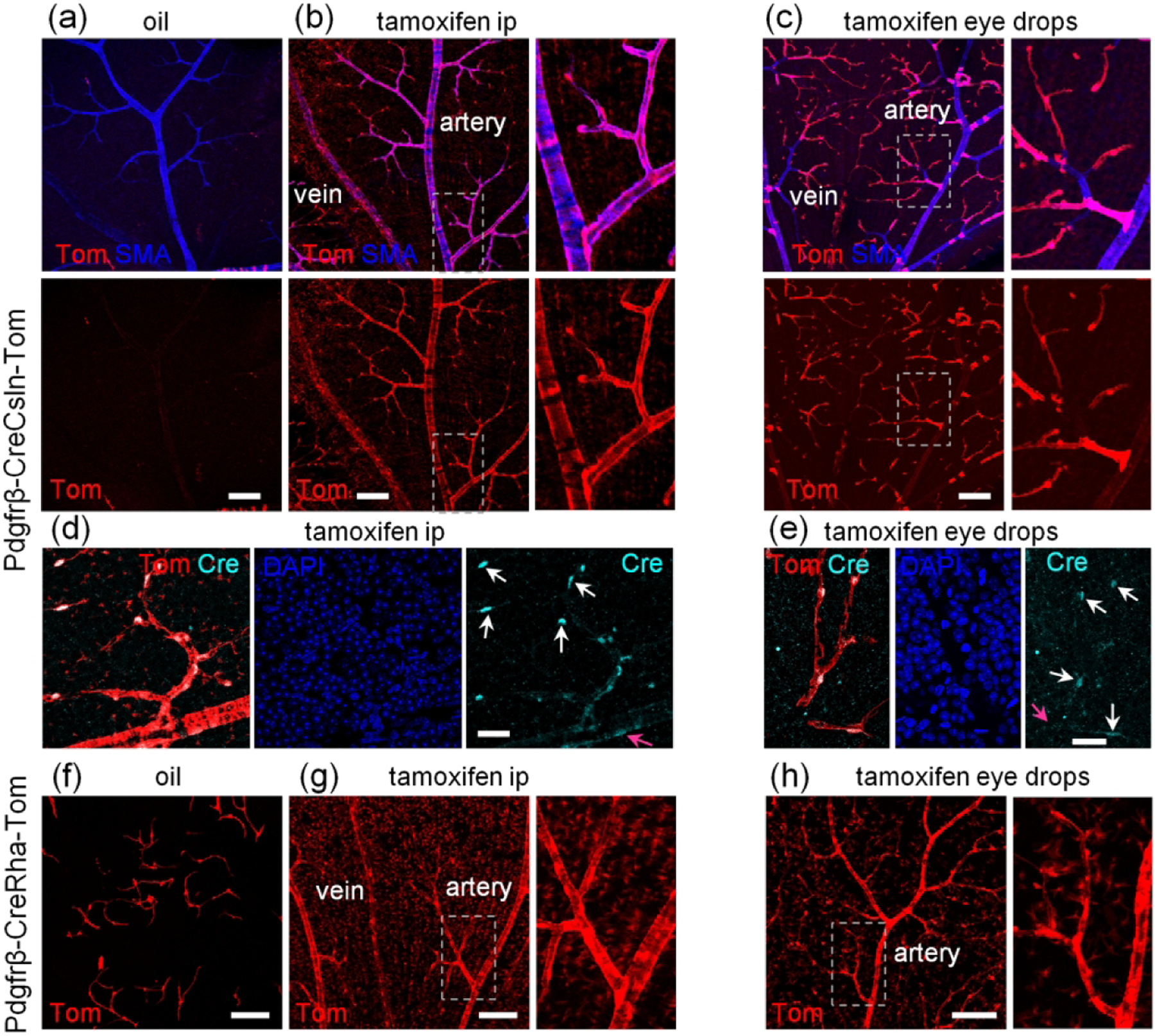
Comparison of two Pdgfrβ-CreERT2 lines for baseline activation of Cre and specificity of Cre activation after tamoxifen induction. (a) Pdgfrβ-CreCsln-Tom line had a very few TdTomato-positive mural cells without tamoxifen induction. (b) After ip tamoxifen treatment, Pdgfrβ-CreCsln-Tom line exhibited prominent TdTomato-labeling in all types of mural cells. All mural cells were positive. Weak TdTomato-expression was detected in Muller cells (magnified region). (c) In Pdgfrβ-CreCsln-Tom treated with tamoxifen eye drops, TdTomato was found predominantly in the capillaries and significantly less frequent in SMA cells and mural cells on veins. (d) After ip tamoxifen treatment, Pdgfrβ-CreCsln-Tom line showed Cre immunoreactivity in mural cell nuclei. The intensity of Cre immunolabeling was higher in pericytes (white arrows) and less in SMA cells (magenta arrows). (e) After tamoxifen eye drop treatment, Pdgfrβ-CreCsln-Tom line showed weak Cre immunoreactivity in pericytes (white arrows). Cre immunolabeling in SMA cells was below detection threshold (magenta arrow). (f) Pdgfrb-CreRha-Tom line had numerous pericytes labeled without tamoxifen treatment. (g) After tamoxifen ip, all mural cells in Pdgfrβ-CreRha-Tom line, were positive for TdTomato; in addition majority of Muller cells were labeled (magnified area). (h) After eye tamoxifen drops, all mural cells were labeled, many Muller cells were also positive. Scale bars: 100 μm in a-c and f-h, 25 μm in d-e.