
FIGURE 4.
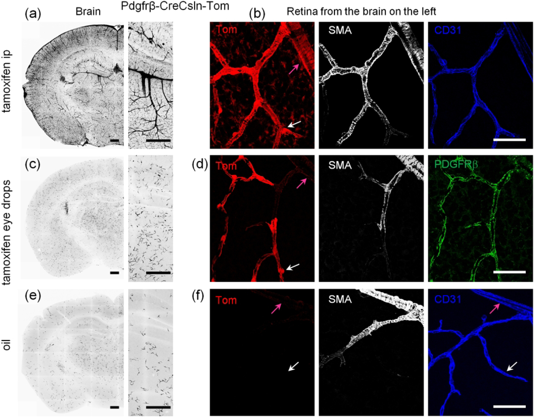
Controlling activation pattern of CreERT2 via tamoxifen administration in B6.Cg-Pdgfrβtm1.1 (cre/ERT2)Csln/J line. (a) In the brain of tamoxifen ip treated Pdgfrβ-CreCsln-Tom all mural cells expressed TdTomato; no glial labeling was visible. (b) In the retina of the mouse from (a), TdTomato-labeling was in SMA cells (magenta arrow) and pericytes (white arrow), as well as many Muller cells. No endothelial cells (blue) were labeled. (c) In the brain of Pdgfrβ-CreCsln-Tom treated with tamoxifen eye drops, TdTomato was found predominantly in the individual separate pericytes in the capillaries but not in large arteries and veins. (d) In the retina of the mouse from (b), TdTomato-labeling was colocalized with Pdgfrb immunolabeling (green) suggesting selective Cre activity in the majority of pericytes (white arrow) but not in SMA cells (magenta arrow). (e) In the brain of oil ip treated Pdgfrβ-CreCsln-Tom, sparse TdTomato labeling was detected in pericytes. (f) In the retina of the mouse from (e), there was no TdTomato-positive cells neither among SMA cells (magenta arrow) nor pericytes (white arrow). Endothelial cell immunolabeling for CD31 was used to visualize capillaries (blue). Scale bars: 400 μm in a, c, and e; 25 μm in b, d, and f.