
Figure 2.
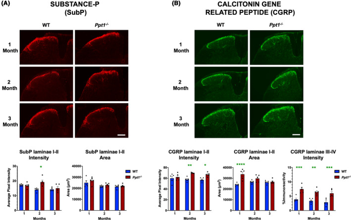
Early alterations in nociceptive markers in Ppt1−/− mouse spinal cords. Immunostaining for (A) Substance‐P (SubP) and (B) calcitonin gene‐related peptide (CGRP) reveal age‐related changes in these markers in the dorsal horn of Ppt1−/− mice. Representative images of dorsal horns of spinal cords reveal an increase in SubP immunoreactivity in laminae I‐II at 2 months and increased CGRP immunoreactivity in laminae I‐II beginning at 2 months as measured by image densitometry (average pixel luminance). Measures of area of immunoreactivity (µm2) confirmed a greater area of CGRP immunoreactivity at 1 month in Ppt1−/− mice across all time points as compared to WT (wild‐type) mice. Thresholding image analysis of positively stained fibres in laminae III‐IV of CGRP‐stained tissue showed a consistent increased in values in Ppt1−/− mice across all time points as compared to WT mice. Scale bars = 200µm. P‐values ‐ *p ≤ 0.05, **p ≤ 0.01, ***p ≤ 0.001, ****p ≤ 0.0001; two‐way ANOVA with post hoc Bonferroni correction. Values shown are mean ± SEM. (n = 5 mice/group).