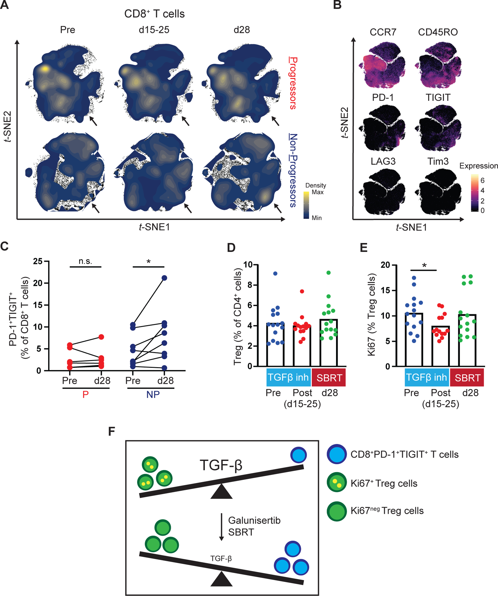
Figure 4. Galunisertib and radiation is associated with changes in T cell subsets in the peripheral blood after treatment.

PBMC from Progressors (P) and Non-Progressors (NP) were analyzed at the indicated timepoints by high-dimensional single cell mass cytometry. Shown are (A) Density plots and (B) Marker expression level plots. (C) Paired comparison of manually gated CD8+PD-1+TIGIT+ cells at baseline (pre) and after combination treatment (d28) in Progressors and Non-Progressors. (D) Quantification of Treg cell (CD25+CD127lowFoxp3+) frequency (percentage of CD3+CD4+) pre-treatment (pre) and post-treatment with galunisertib (d15–25) and SBRT (d28). Cell subsets were gated after the exclusion of doublets and dead cells and positive selection for CD45, CD3 and CD4. (E) Quantification of Ki67+Treg cells (percentage of Treg cells) in samples pre-treatment and post-treatment with galunisertib and SBRT. (F) Schematic detailing the proposed association between TGF-β signaling and relevant peripheral blood leukocytes. At baseline Ki67+Tregs and low frequencies of CD8+PD-1+TIGIT+ T cells are found in the peripheral blood. After treatment, in non-progressors, the peripheral blood leukocyte composition is shifted towards higher frequencies of CD8+PD-1+TIGIT+ T cells and lower frequencies of Ki67+Tregs. The size of TGF-β lettering indicates higher (large) or lower (small) TGF-β signaling activity. Wilcoxon matched-pairs tests were performed (C). Mann-Whitney tests were performed (D-E). *, p < 0.05. SBRT, stereotactic body radiation therapy.
