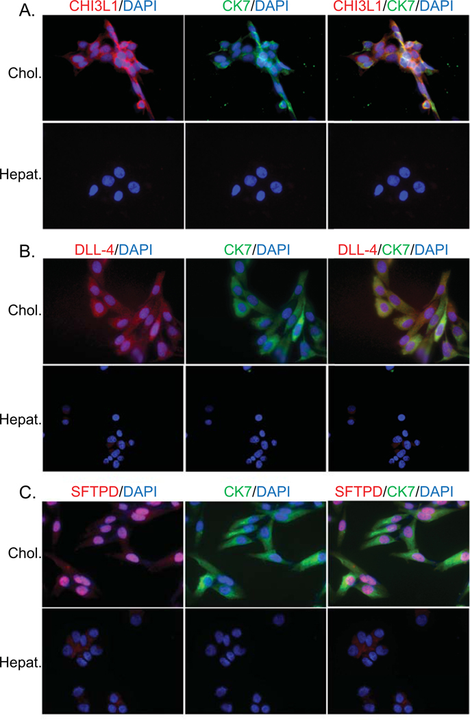
Figure 4. Cholangiocyte-predominant localization of candidate autoantigens.
Immunofluorescence of hepatocyte (Hepat.) or cholangiocyte (Chol.) cell lines with fluorochrome-tagged antibodies to cytokeratin 7 (CK7- cholangiocyte specific; green), CHI3L1 (A.), DLL-4 (B.) or SFTPD (C.) (all autoantigens in red) and counterstained with nuclear DAPI (blue). Co-localization of CK7 and an autoantibody is reflected in yellow color.

