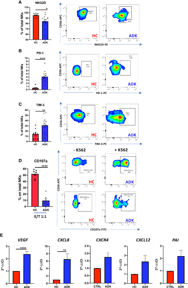
Figure 2.
pNK cell exhaustion and degranulation activities in peripheral blood of PCa patients. PCa pTA-NKs have decreased levels of the NKG2D activation markers (A), increased expression of the PD-1 (B) and TIM-3 (C) exhaustion markers and impaired degranulation abilities against the K562 cells (D). panel D shows NK cell degranulation capabilities, alone or co-incubated with K562 cells in PCa p-TA-NKs and NK cell from healthy controls. Every dot in dots/bars graph refers to single patient or control. Representative dot plots show the specific antigen expression (as % of total pNK cells). pNK cells, FACS-sorted from patients with ADK-PCa have increased expression of the pro-inflammatory factors VEGF, CXCL8, CXCR4, CXCL12, PAI (E). qPCR have been performed using pNK cell from 3 PCa patients and 3 controls, in triplicate. Data are showed as mean ± SEM, t-student test, *p<0.05, **p<0.01, ****p<0.0001. HC, healthy controls; ADK, prostate cancer adenocarcinoma.

