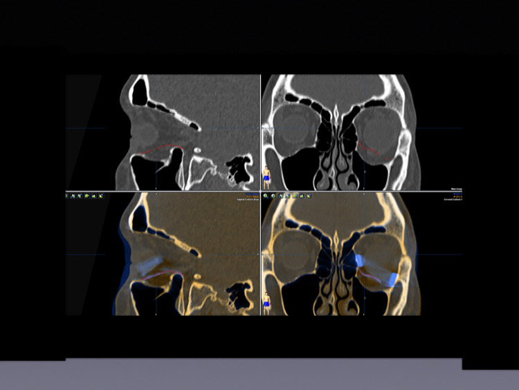
Figure 3.
Planning of implant placement and volume augmentation using titanium spacers. Upper left: Sagittal view—SLM implant (red) placed to reconstruct the orbital floor. Upper right: Axial view of the placed SLM implant (red). Lower left: Sagittal view of the SLM implant (red) with an additional titanium spacer (blue) used to augment volume. Lower right: Coronal view of the midface. SLM implant (red) placed on the left orbital floor and two titanium spacers placed at the lateral and the medial lower orbital walls to add volume.

