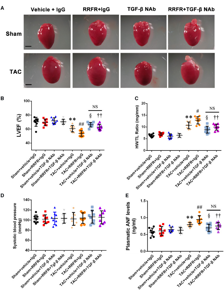
Figure 6.
The RRFR peptide accelerates cardiac hypertrophy. (A) Morphology of the hearts from sham mice and TAC mice injected with the RRFR peptide in combination with or without TGF-β NAb for 8 weeks. Scale bar = 1 mm. (B) Echocardiographic data showing left ventricular ejection fractions (LVEF, n = 8/group). (C) Ratio of heart weight to tibia length (HW/TL, n = 8/group). (D) Systolic blood pressure (mmHg, n = 8/group). (E) Plasma ANF levels (n = 8/group). Data are shown as mean ± SD. **P < 0.01 vs. Sham+vehicle+IgG group; #P < 0.05, ##P < 0.01 vs. TAC+vehicle+IgG group; §P < 0.05, vs. TAC+vehicle+IgG group; ††P < 0.01 vs. TAC+RRFR+IgG group. NS, not significant. Statistical analysis was carried out by a one-way ANOVA test.

