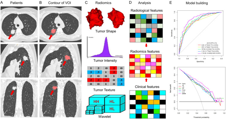
Figure 1.
Radiomics features extraction and analysis workflow. A. Original CT images of a patient with lung adenocarcinoma. B. Segmentation of the tumor volume of interest (VOI) on all CT slices by experienced radiologists. C. Feature extraction from the VOI, including tumor shape, intensity, texture, and wavelet features. D. Clinical, radiological, and radiomics feature analysis. E. Model building.

