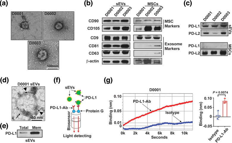
FIGURE 1.

PD‐L1 is enriched on WJMSC‐derived sEVs. (a) Representative TEM imaging of purified sEVs from three clinical grade human WJMSCs (D0001, D0002 and D0003). Scale bar, 100 nm. (b‐c) Immunoblotting for stem cell biomarkers and exosome biomarkers (b), and checkpoint PD‐L1 and PD‐L2 (c) in purified WJMSC sEVs and the whole cell lysates (MSCs). All lanes were loaded with the same amount of protein. (d) A representative TEM image showing the immunogold‐labelled PD‐L1 signals (arrowheads) on the surface of a WJMSC sEV (arrow). Scale bar, 50 nm. (e) Immunoblotting for PD‐L1 in the whole exosome lysates (Total) and exosomal membrane protein extracts (Mem). All lanes were loaded with the same amount of protein. (f) Schematic of optical biolayer interferometry (BLI) assay for detecting surface PD‐L1 on the WJMSC sEVs. PD‐L1‐Ab, PD‐L1 detection antibody. (g) A typical binding (left) between PD‐L1‐Ab and WJMSC sEVs (red curve) or between Isotype and WJMSC sEVs (blue curve) and quantification analysis based on their wavelength differences (right, n = 3). Data are mean ± s.e.m. and analysed by unpaired one‐tailed Student's t‐test
