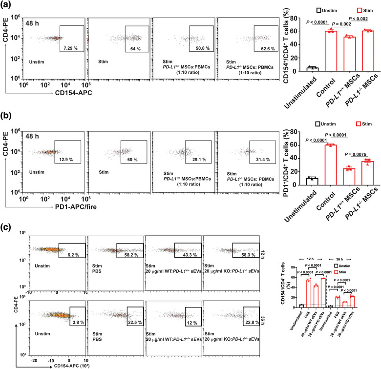
FIGURE 3.

Both WJMSCs and sEVs decrease their capability to block TCR‐mediated TCA after PD‐L1 knockout. (a) Flow cytometry showing the 48‐hour inhibitory effects of both WT and KO WJMSCs on CD4+ T cell activation (left) and quantitative analysis (right). (b) Expression of PD1 on the CD4+ T cells mentioned above, measured by flow cytometry (left) and quantitation (right). (c) Representative flow charts (left) and quantitative analysis (right) of activated CD4+ T cells incubated with 20 µg/ml PD‐L1 WT or KO WJMSC sEVs for 12 h or 36 h. PBMCs were stimulated with (Unstim) or without (Stim) CD3/CD28 Dynabead at a dilution of 1:1 ratio (a‐c) and the ratio of WJMSCs to PBMCs was 1 to 10 (a,b). Data are mean ± s.e.m (n = 3) and analysed by one‐way ANOVA (a‐c). Data are representative of three independent experiments (a‐c)
