Figure 5.

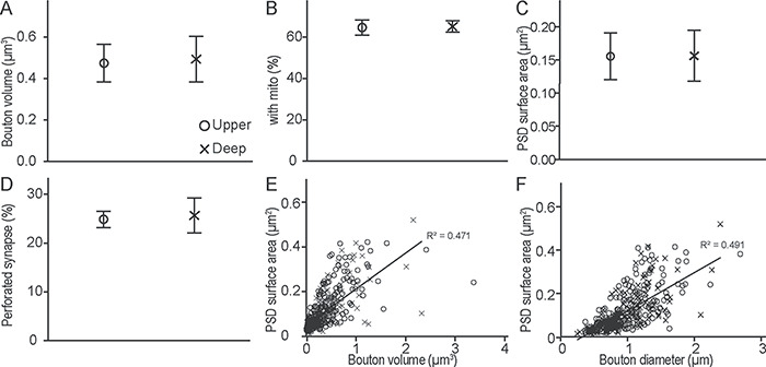
Presynaptic and postsynaptic features of hippocampal boutons in A25. (A) Hippocampal bouton volume was similar in the upper and deep layers of A25. (B) Proportion of hippocampal boutons that contained mitochondria (mito) and formed synapses in the upper and deep layers of A25. (C) PSD surface area of postsynaptic sites that receive hippocampal projections in the upper and deep layers of A25. (D) Proportion of hippocampal boutons that formed perforated synapses in the upper and deep layers of A25. (E, F) Relationship of PSD surface area and bouton volume (E) or diameter (F) for all cases. Upper layers, circles; deep layers, crosses. Error bars: ±SE.
