Figure 8.

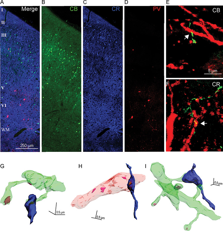
Relationship of hippocampal terminations to inhibitory neurons in the upper and deep layers of A25. (A–D) Immunofluorescence photomicrographs of three adjacent sections of A25 labeled with CB (green), CR (blue), and PV (red). (A) Superimposed sections (B–D) show all three neurochemical classes of inhibitory neurons. Scale bar, 250 μm. (E–F) Immunofluorescence photomicrographs show apposition sites between hippocampal terminations (green) and CB (E) or CR (F) positive elements (red); white arrows indicate apposition sites. Scale bar, 5 μm. Error bars: ±SE. (G–I) 3D reconstruction of labeled hippocampal boutons (blue) and their postsynaptic targets. (G) A hippocampal bouton forming a synapse on a spine (green). (H) A hippocampal bouton forming a synapse on an aspiny dendritic shaft (pink) of a presumed inhibitory neuron. (I) A hippocampal bouton forming a synapse on two spines that come from two distinct dendritic shafts. Color codes: green, spiny dendrites; pink, aspiny dendrite of a presumed inhibitory neuron; yellow–brown, PSD; magenta, PSD by unlabeled postsynaptic sites on dendrite; scale bar: 0.5 × 0.5 × 0.5 μm.
