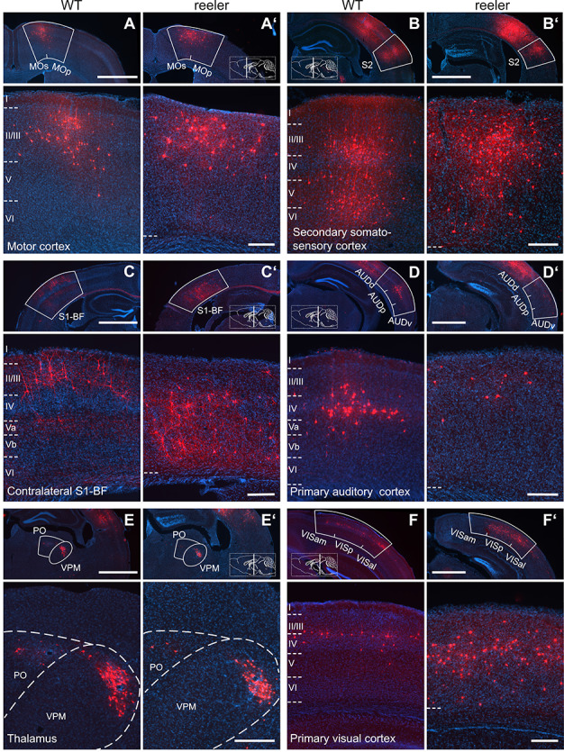
Figure 3.

Long-range input to VIP cells in barrel cortex of WT and reeler mice. (A/A’–F/F’) Sections along the rostrocaudal extent of WT and reeler mice showing consistently labeled areas with presynaptic partners of VIP cells in barrel cortex. In the top overview panels, the white contour delineates the borders of the respective source area. Higher magnification close-ups are shown below. Section planes are indicated on the schematic sagittal brain section (scale bar overview: 1000 μm; scale bar close-up: 200 μm; AUDd/AUDp/AUDv, dorsal/primary/ventral auditory area; MOp/MOs, primary/secondary motor cortex; PO, posterior complex of the thalamus; S1-BF, primary somatosensory cortex, barrel field; S2, secondary somatosensory cortex; VISal/VISam/VISp, anterolateral/anteromedial/primary visual area; VPM, ventral posteromedial nucleus of the thalamus).
