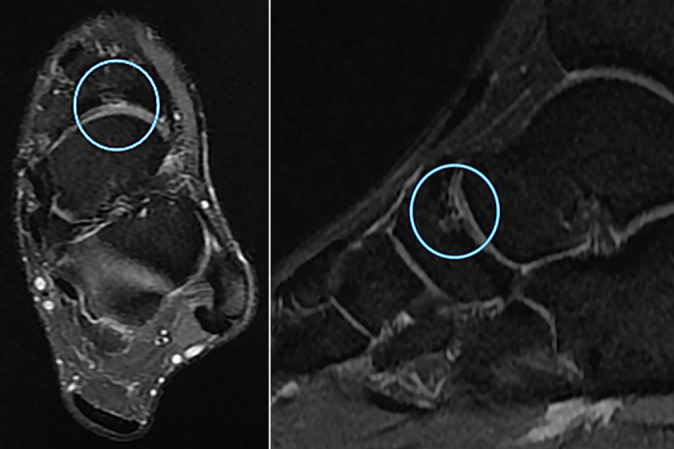
Figure 5.

Sagittal and axial magnetic resonance imaging scans of a cartilage lesion of the transverse tarsal joint in a 21-year-old professional soccer athlete. Signs of grade 3 (modified Noyes and Stabler classification) cartilage lesion at the level of the transverse tarsal (Chopart) joint (in circle) in the form of thinning of the articular cartilage with exposure of the articular surfaces and signs of bone marrow edema in the subchondral navicular bone.
