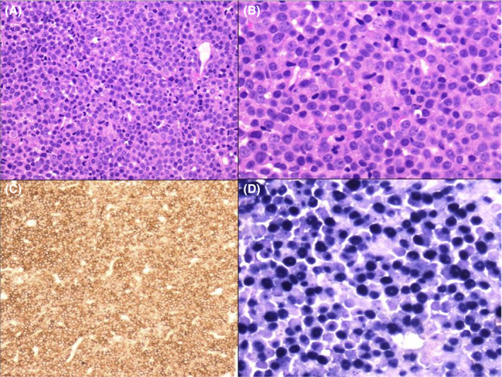
Figure 1.

(A) Histologic section of breast biopsy showing diffuse infiltration by sheets of plasmablasts (H&E × 100). (B) Higher power view of the same area (H&E × 400). (C) Diffuse positivity of plasmablasts with CD138 immunohistochemistry (×100). (D) In situ hybridization for Epstein‐Barr virus‐encoded RNA (EBER) shows positive staining in approximately 99% of plasmablasts (blue nuclear positivity) (×400)
