
Figure 2.
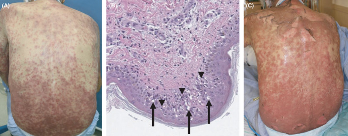
Skin photograph and histological image at the onset of Stevens‐Johnson syndrome (SJS)/ Toxic epidermal necrolysis (TEN). A, Skin photograph at the onset of SJS. B, Necrotic changes in epidermal keratinocytes, vacuolar degeneration in the epidermal layer (arrow), and lymphocyte infiltration into the epidermal layer (arrow head). C, Skin photograph at the onset of TEN