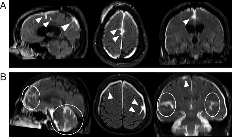
Figure 4.
Multiplanar views of extravasation of contrast into subarachnoid space (ECSAS), representing a breach of the blood-subarachnoid barrier. (A) A subject’s delayed post-contrast FLAIR shows ECSAS in the falx, with sagittal, axial and coronal views (left to right) clearly demonstrating the three-dimensional extravasation of contrast. (B) ECSAS can also occur along the bilateral convexities and cerebellum, as seen in another subject’s delayed post-contrast FLAIR. The white arrowheads and ellipses indicate areas of ECSAS.

