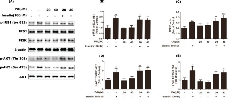
Fig. 5.
Effects of PA on insulin-stimulated phosphorylation of IRS1 and Akt, and protein expression of PI3K in C2C12 myotubes. Cells were incubated with PA (20 and 40 µM), insulin (100 nM), or PA and insulin for 24 h. Western blotting of whole cell lysates to detect phosphorylation (p) of IRS1 and Akt, and protein expression of PI3K. Western blotting (a) and quantification (b–e) of phospho-IRS1, phospho-Akt, and PI3K (n = 3 or 4). Values are expressed as mean ± SD. * P < 0.05, ** P < 0.01, versus control group.

