Figure 1.

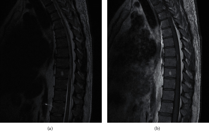
(a) Sagittal T2 MRI of dorsal spine showing high T2 signal with cord edema from T4 to T9 levels with thickened epidural fat tissue posteriorly. (b) Sagittal T1 MRI with gadolinium of dorsal spine showing an enhancing lesion at T7-T8 level.

(a) Sagittal T2 MRI of dorsal spine showing high T2 signal with cord edema from T4 to T9 levels with thickened epidural fat tissue posteriorly. (b) Sagittal T1 MRI with gadolinium of dorsal spine showing an enhancing lesion at T7-T8 level.