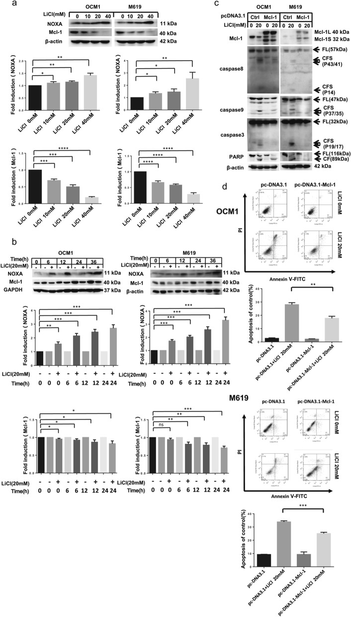
Fig. 4.
The NOXA/Mcl-1 axis contributes to LiCl-induced apoptosis. a OCM1 and M619 cells were treated with 0, 10, 20, or 40 mM LiCl for 36 h and then harvested for western blotting analysis. b OCM1 and M619 cells were treated with 20 mM LiCl for 0, 6, 12, 24 and 36 h and then harvested for western blotting analysis. In order to make the comparison more concise and observe the cell, we added a control for 0 h, 6 h, 12 h and 24 h. NOXA and Mcl-1 expression was quantified using ImageJ software and analysed with GraphPad Prism 5.0 software. c d OCM1 and M619 cells were seeded in 6-well plates and transfected with vehicle-treated or pc-DNA3.1- Mcl-1 plasmids on the second day. After 48 h of transfection, the cells were exposed to 20 mM LiCl for 24 h and then harvested for western blotting c and apoptosis analysis d. All data are presented as the mean ± S.D. *P < 0.05, **P < 0.01, ***P < 0.001, ****P < 0.0001

