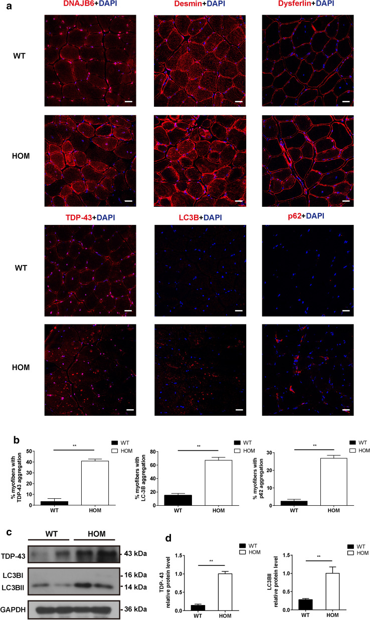
Fig. 7.
Aggregation of myofibril components in skeletal muscle tissue of 18-month-old HOM and WT mice. a DNAJB6, desmin, dysferlin, TDP-43, LC3B and p62 staining displayed altered subcellular distribution in the HOM mice. Scale bar = 20 µm. b Percentage of myofibers with TDP-43, LC3B and p62 aggregation in 18-month-old HOM and WT mice. c Western blot showing TDP-43 and LC3B in muscle lysates of 18-month-old HOM and WT mice. GAPDH was used as a loading control. d Quantification of relative intensities of proteins shown in c. Data represent means ± SEM of 3 independent experiments (n = 3). P value = * < 0.05, ** < 0.01

