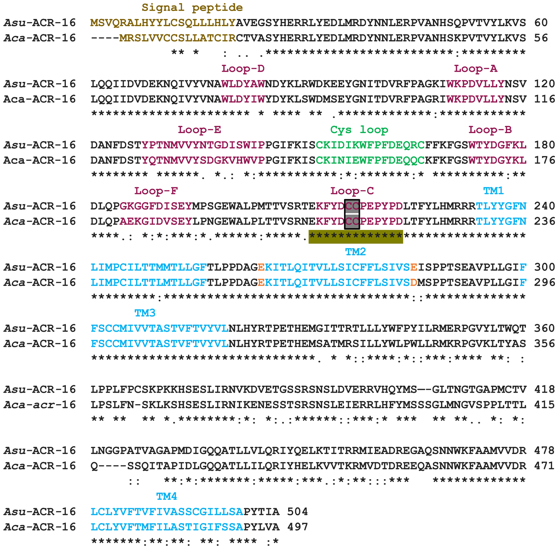
Fig. 1.

Amino acid sequence alignment of Aca-ACR-16 and Asu-ACR-16. The signal peptide (light brown), ligand binding loops (A to F; maroon) transmembrane regions TM1–4 (blue) and cys-loop (green) are indicated. The adjacent cysteines (in the Y–x–C–C motif) in loop-C are indicated in the black box. The negatively charged amino acids (E: Glutamic acid and D: Aspartic acid) flanking the TM2 domain are highlighted in orange. Residues involved in binding of α-BTX are highlighted in olive green. Note: The sequence of Aca-ACR-16 amplified from A. caninum larval total RNA is shorter than the WormBase sequence ANCCAN_01899 and lacks 19 amino acids (KVKEPNLFGPWENFHGDLF) between the cys-loop and loop-B. These amino acid residues are also lacking in the A. suum ACR-16 homologue
