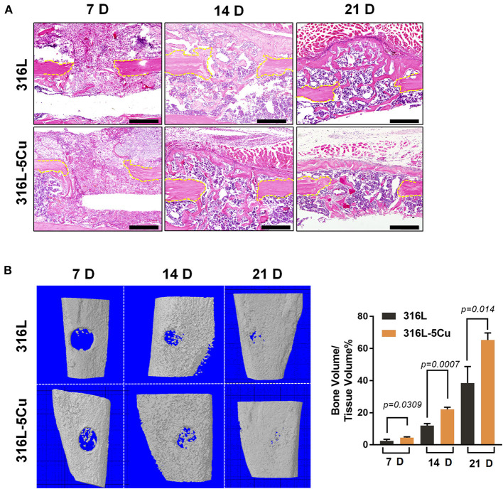
Figure 2.
Copper-containing metal (CCM) accelerated callus formation in bone repair. (A) HandE-stained histological images of the tibia of 316L/316L−5Cu-inserted mice 7, 14, and 21 days after injury. Yellow dotted lines indicate the edge of the drill hole. Scale bar represents 200 μm. (B) Three-dimensional microcomputed tomography (μCT) images of the drill holes in 316L/316L−5Cu-inserted mice 7, 14, and 21 days after injury. The ratio of bone volume to tissue volume (BV/TV), representing bone formation in the drill holes, was calculated. Data were statistically analyzed by Student's t-test. Results are presented as mean ± SEM. n = 6 per time point per group.

