Figure 5.

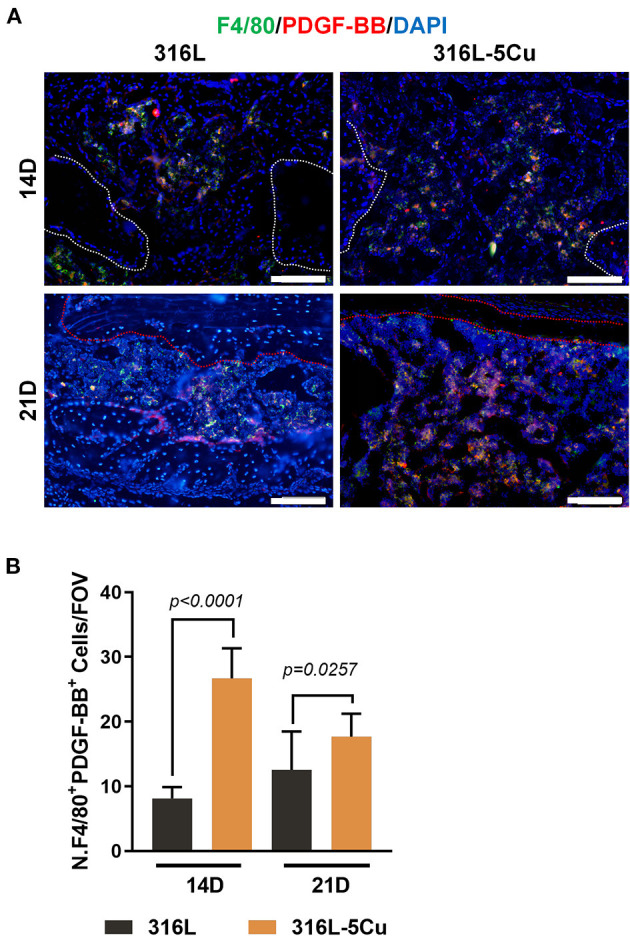
M2a macrophage-derived platelet-derived growth factor type BB (PDGF-BB) levels were elevated by 316L−5Cu. (A) Representative images of double-immunofluorescence staining against F4/80 (green) and PDGF-BB (red) at days 14 and 21. The merged yellow shows colocalization of F4/80 and PDGF-BB. Scale bars represent 100 μm. (B) Quantification of numbers of F4/80+ and PDGF-BB+ cells within drill holes in 316L/316L−5Cu inserted mice at days 14 and 21. Data were collected from at least three fields of view per sample and six samples per group. Data were statistically analyzed by Student's t-test. Results are presented as mean ± SEM.
