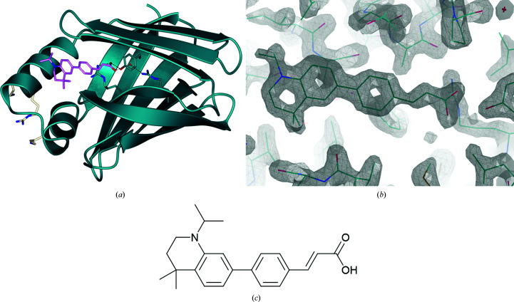
Figure 3.
(a) DC479 (depicted in stick representation, pink) incorporated into the binding site of CRABPII (ribbon representation, dark blue; O atoms, red; N atoms, blue). The key binding resides (Arg112-Arg133-Tyr135) and a conserved water molecule can be seen at the base of the hydrophobic pocket with predicted hydrogen bonds to the ligand (dark blue; O atoms, red; N atoms, blue). NLS-forming residues (white; N atoms, blue) can be seen facing externally on the left. The protein forms a dimer with noncrystallographic symmetry, of which chain A is displayed for clarity. The r.m.s.d. between chains A and B on Cα atoms is 0.23 Å. (b) Ligand density of DC479 in chain A in the ligand-binding site of CRABPI (2F o − F c map including ligand, contour σ = 1.50). (c) Chemical structure of DC479.

