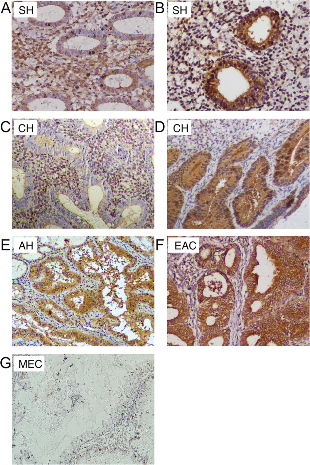
Figure 1.

The expression pattern of PRMT5 in EC and a series of endometrial hyperplasia. (A) PRMT5 was mainly expressed in stromal cells and in the cytoplasm and nuclei of some glandular cells in some cases of SH; (B) a PRMT5‐positive case of SH (both glandular cytoplasmic and nuclear positivity, with stromal cell positivity); (C) PRMT5 was mainly expressed in stromal cells in some cases of CH; (D) PRMT5‐positive case of CH (both glandular nuclear and cytoplasmic positivity); (E) PRMT5‐positive case of AH (moderate intensity cytoplasmic positivity, with a small number of positive nuclei); (F) PRMT5 expression showed strong cytoplasmic staining and limited nuclear localization in EAC; (G) PRMT5 was barely expressed in MEC. AH, atypical hyperplasia; CH, complex hyperplasia; EAC, endometrioid adenocarcinoma; MEC, mucinous endometrial carcinoma; SH, simple hyperplasia.
