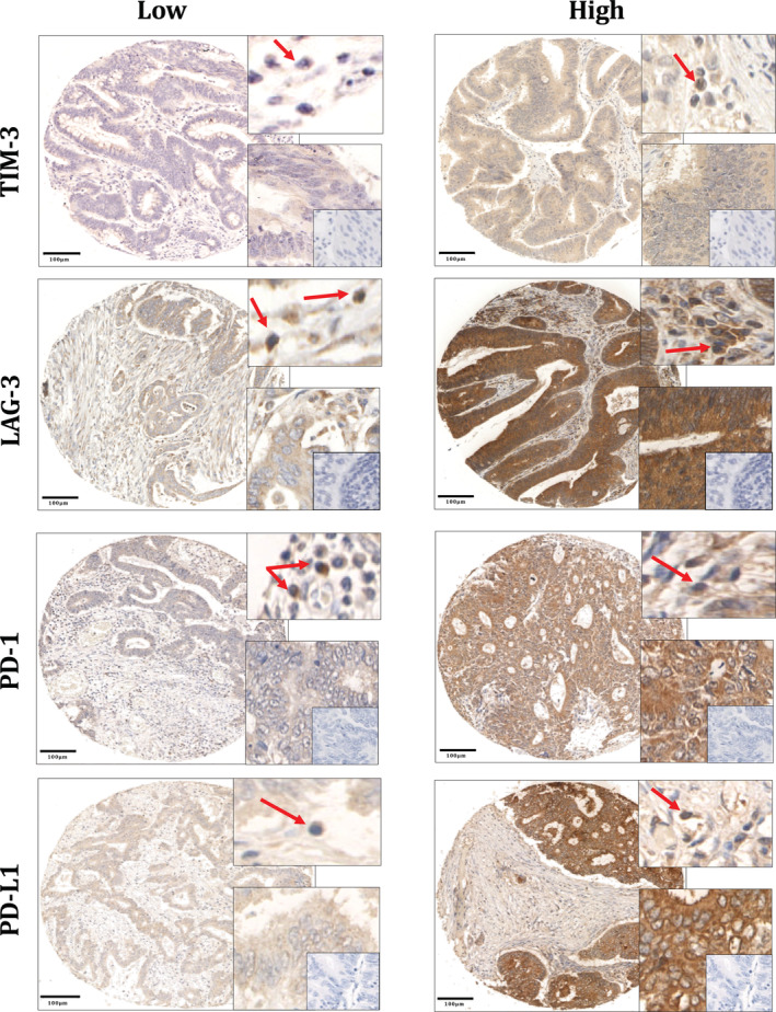
Figure 1.

Immune checkpoint expression in colorectal cancer. TIM‐3, LAG‐3, PD‐1 and PD‐L1 showed cytoplasmic staining. For each marker, the top right boxes show examples of positive lymphocytes in the stroma (red arrows); and the bottom right boxes show positive tumour cells at a higher magnification with no‐antibody negative controls within (insets). Scale bars at 100 μm.
