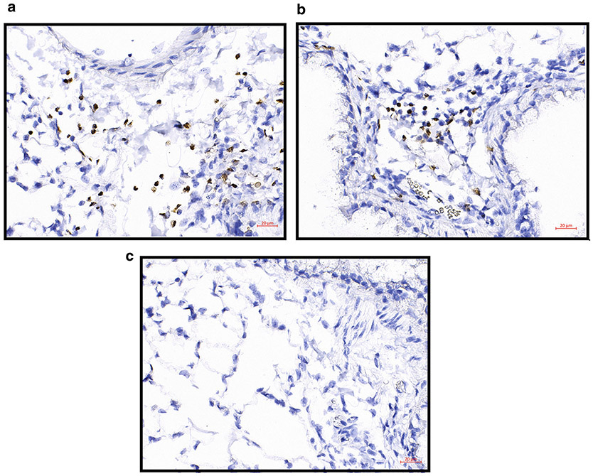
Fig. 6.
Allergen-challenged FFPE lung sections with EPX IHC with DAB as the chromogen. (a, b) Two examples of EPX IHC in allergen-challenged lung FFPE slices. EPX is stained brown showing the location of eosinophils, and hematoxylin counterstains nuclei blue/purple. (c) Negative control staining. Images were taken on Zeiss Imager.M2 with a ×40 objective

