Fig. 3.

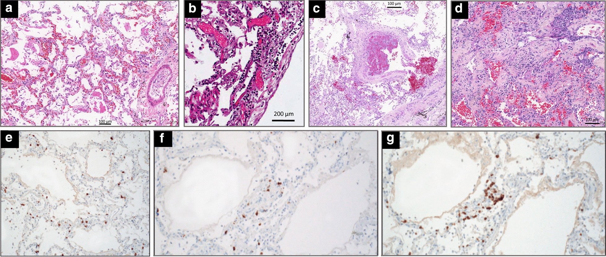
Microscopic findings in the lungs of five patients who died from coronavirus disease 2019. a In a 76-year-old man with hypertension who died from a cardiac arrest 10 days after the onset of symptoms (no admission in the ICU), diffuse alveolar damage with vascular congestion, oedema and perivascular lymphocytic infiltration is present; b In a 78-year-old man with hypertension, morbid obesity, diabetes type 2 who died from hypoxic cardiorespiratory failure three days after hospital admission and two days of non-invasive ventilation, interstitial pneumonia with perivascular lymphocytic infiltration of interalveolar septa and multifocal endothelialitis is present; c In a 63-year-old man without co-morbidity who died from hypoxic cardiorespiratory failure 37 days after onset of symptoms and 26 days after ICU admission and invasive ventilation, partial thrombosis of a pulmonary arteriole with perivascular lymphocytic infiltration is present; d In a 64-year-old man without co-morbidity who died from hypoxic cardiorespiratory failure 21 days after onset of symptoms and 15 days after ICU admission and invasive ventilation, diffuse alveolar damage at a proliferative phase is present with collagen plugs deposition in alveolar spaces (hematoxylin staining collagen in light pink and paucicellular areas); e–g In a 77-year-old man with hypertension and mild obesity, who died on hospital admission six days after onset of symptoms, an interstitial pneumonia is present. T-lymphocytes are highlighted by immunohistochemical stains for CD3 (e), CD4 (f), and CD8 (g).
Reproduced from the Department of Pathology of Hospital das Clinicas, Sao Paulo, Brazil (a, c and d) and Permission was granted by Ackermann et al. (©Massachusetts Medical Society [29]) to reuse this figure (b) and Permission was granted by Barton et al. (©Oxford University Press [31]) to reuse this figure (e–f)
