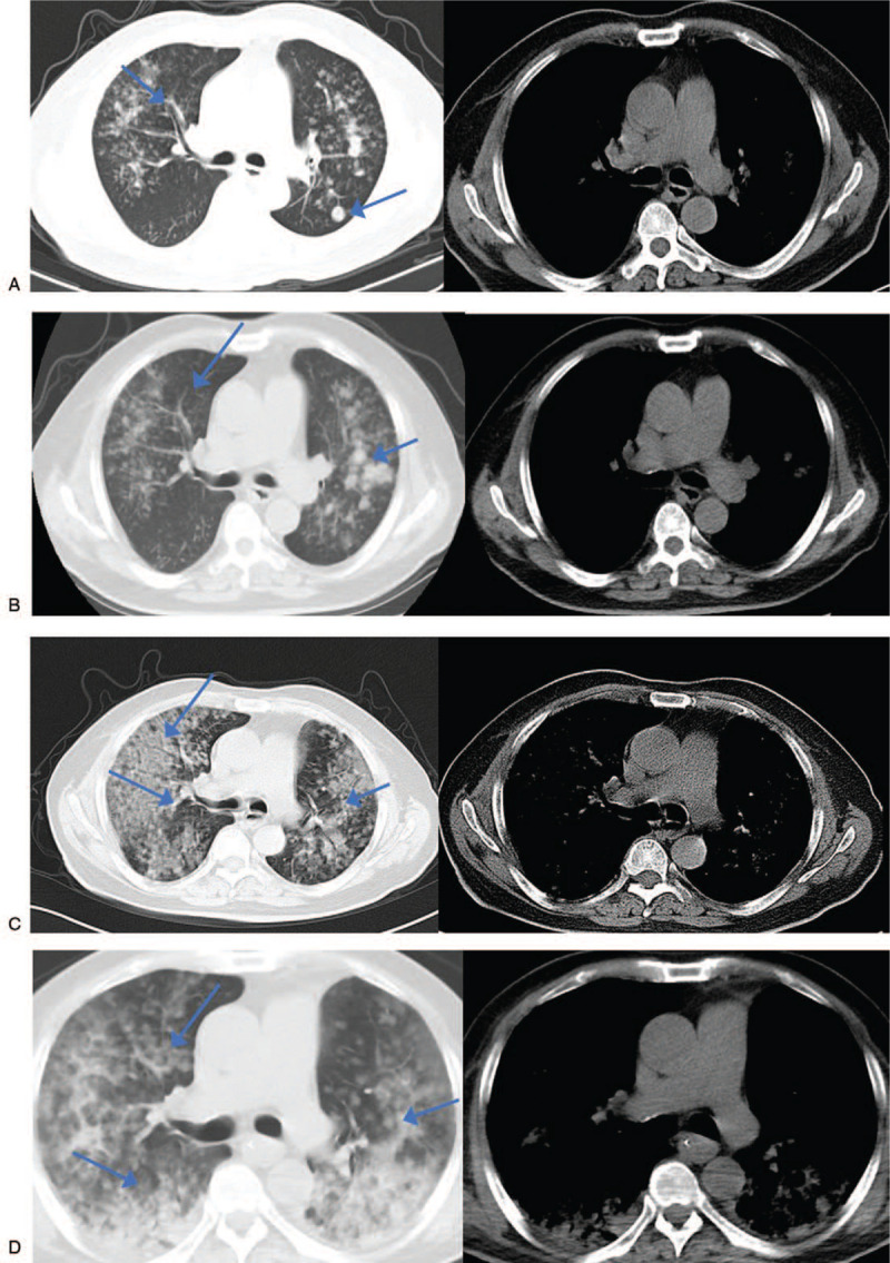
Figure 1.

A. January 19, 2020 chest CT indicating a special infectious lesion that needed to be checked and a nodular shadow in the back of the left lower lobe. B. January 26, 2020 chest CT reexamination indicated diffuse lesions of both lungs, scattered ground glass nodes in both lungs and nodular shadow in the posterior segment of the upper lobe of the left lung. The nodular shadow in the back of the left lower lobe had disappeared. C. April 24, 2020 chest CT reexamination indicating diffuse lesions of both lungs and a special pathological bacterial infection lesion that needed to be examined. Compared with the film on January 26, 2020 the foci had increased significantly and additional diagnostic tests were suggested. Scattered ground glass nodes with a withered shape were seen after careful film-reading, which was obviously more severe than the one on January 26. However, the nodular shadow in the posterior segment of the upper lobe of left lung had absorbed slightly. D. May 14, 2020 chest CT reexamination indicating diffuse lesions of both lungs and the special pathological bacterial infection lesion that needed to be examined. Compared with the film on February 24, 2020 the foci in the upper lobe of the right lung was reduced while the one in the lower lobe of both lungs was increased. Scattered ground glass nodes with a withered sign were seen after careful film-reading. CT = computed tomography.
