
Figure 1.
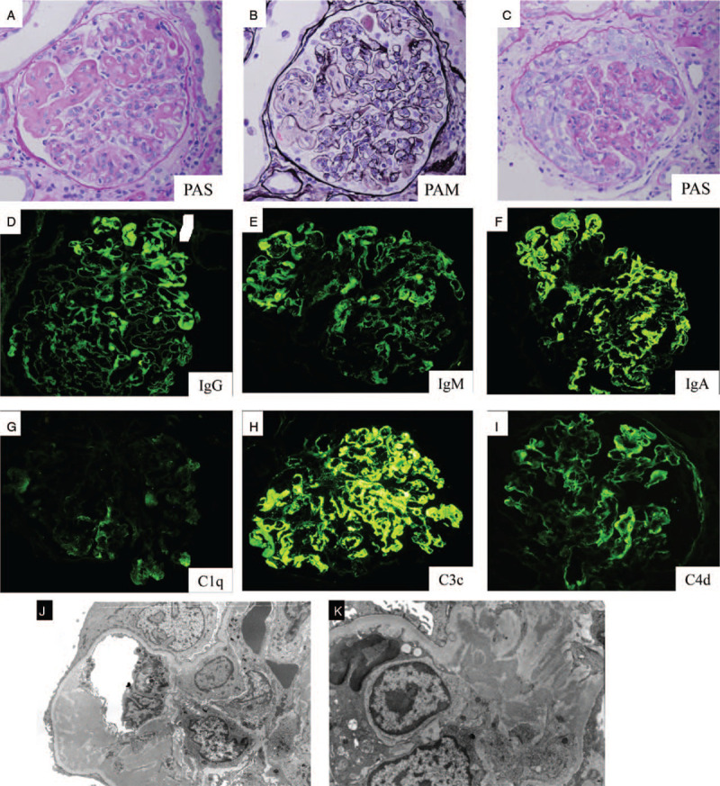
Light microscopy shows staining with periodic acid-Schiff (PAS) and periodic acid–methenamine silver stain (PAM). Original magnification × 200 (A, B) Glomerulus exhibits massive subendothelial deposits, such as wire loop lesion and endocapillary proliferation. (C) Glomerulus displays cellular crescents and endocapillary proliferation. Immunofluorescence of kidney biopsy shows staining with (D) IgG, (E) IgM, (F) IgA, (G) C1q, (H) C3c, (I) C4d. Original magnification × 200. Electron microscopy shows mesangial and endothelial unorganized electron dense deposit, in (J) low and (K) high magnification.