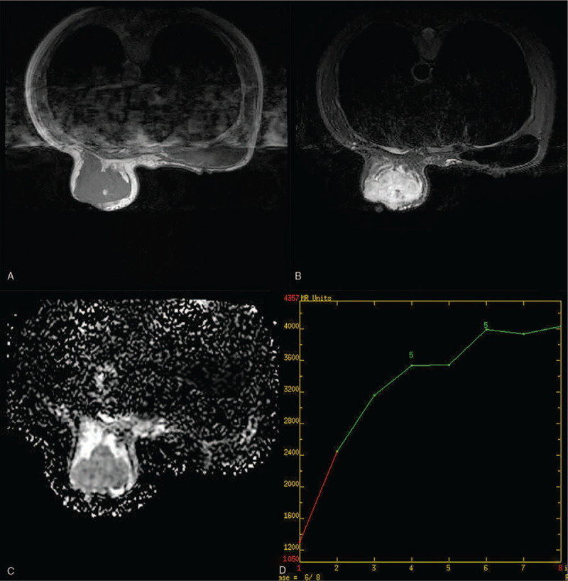
Figure 3.

Breast magnetic resonance imaging of a 47-year-old woman with primary breast angiosarcoma of the left breast. (A) Hypointense with scattered mixed high signal intensity foci on T1-weighted images. (B) Hyperintense on T2-weighted images. (C) High apparent diffusion coefficient value. (D) Dynamic contrast-enhanced imaging with time signal intensity curves for type 1.
