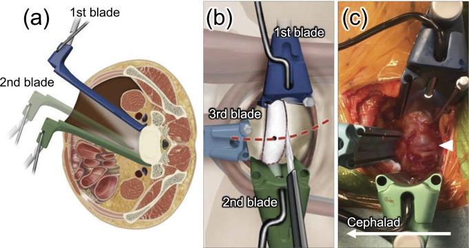
Figure 4.
Exposure of the L5-S1 disc. (a) Placement of 1st and 2nd blades by laterally retracting the common iliac vessels. (b) The L5-S1 disc exposed using three retractor blades. The 1st and the 3rd blades are fixed using pins. The red dot line indicates the midline of the L5-S1 disc. (c) Intraoperative images of the L5-S1 disc. Only the 1st retractor is fixed to the sacrum. White arrowhead indicates the medial sacral vessels.

