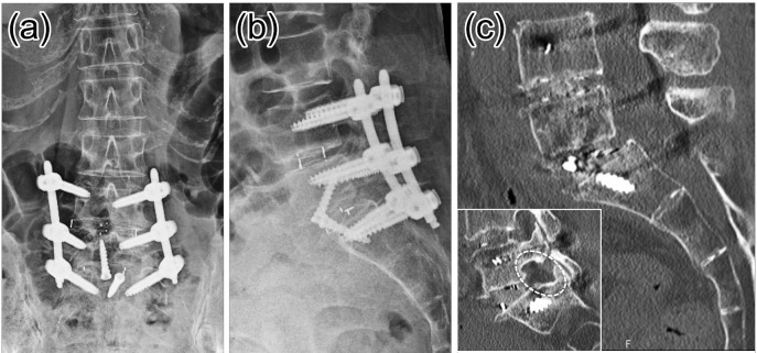
Figure 8.
The patients underwent OLIF51 surgery combined with L4-5 OLIF, followed by posterior pedicle screw fixation using single position procedure with no patient flipping to a prone position. (a, b): Plain X-ray (c) Sagittal and parasagittal CT reconstruction. Dotted circle indicates the impaired left L5-S1 foramen, and note that the foraminal area enlarged compared with the Fig. 7 (c).

