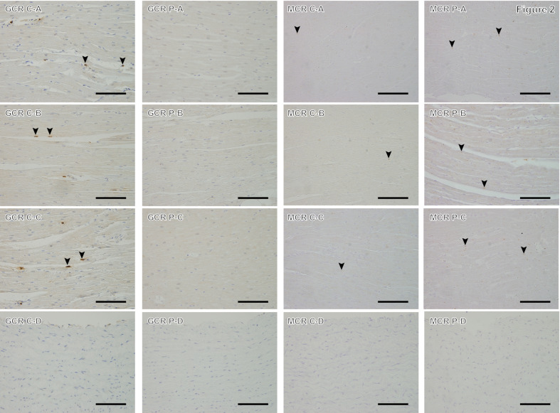
Fig. 2.
Immunostaining of glucocorticoid receptor and mineralocorticoid receptor in the control group (C) and high-dose prednisolone group (P). A: Myocardium of the left ventricular wall; B: myocardium of the right ventricular wall; C: interventricular septum; and D: aorta; GCR: anti-glucocorticoid receptor immunostaining; MCR: anti-mineralocorticoid receptor immunostaining. GCR sections in the C group show brown-stained GCR-positive nuclei (arrowheads) and violet-stained negative nuclei. P group does not show any GCR-positive nuclei in the shown fields. MCR sections in the C and P groups’ myocardium shows brown-stained MCR immunoreactivity positive points (arrowheads) and violet-stained negative nuclei, whereas there are fewer MCR-positive staining points in group C. For the aorta, both groups showed less immunoreactivity for GCR and MCR. Magnification: 400×, scale bar=50 µm.

