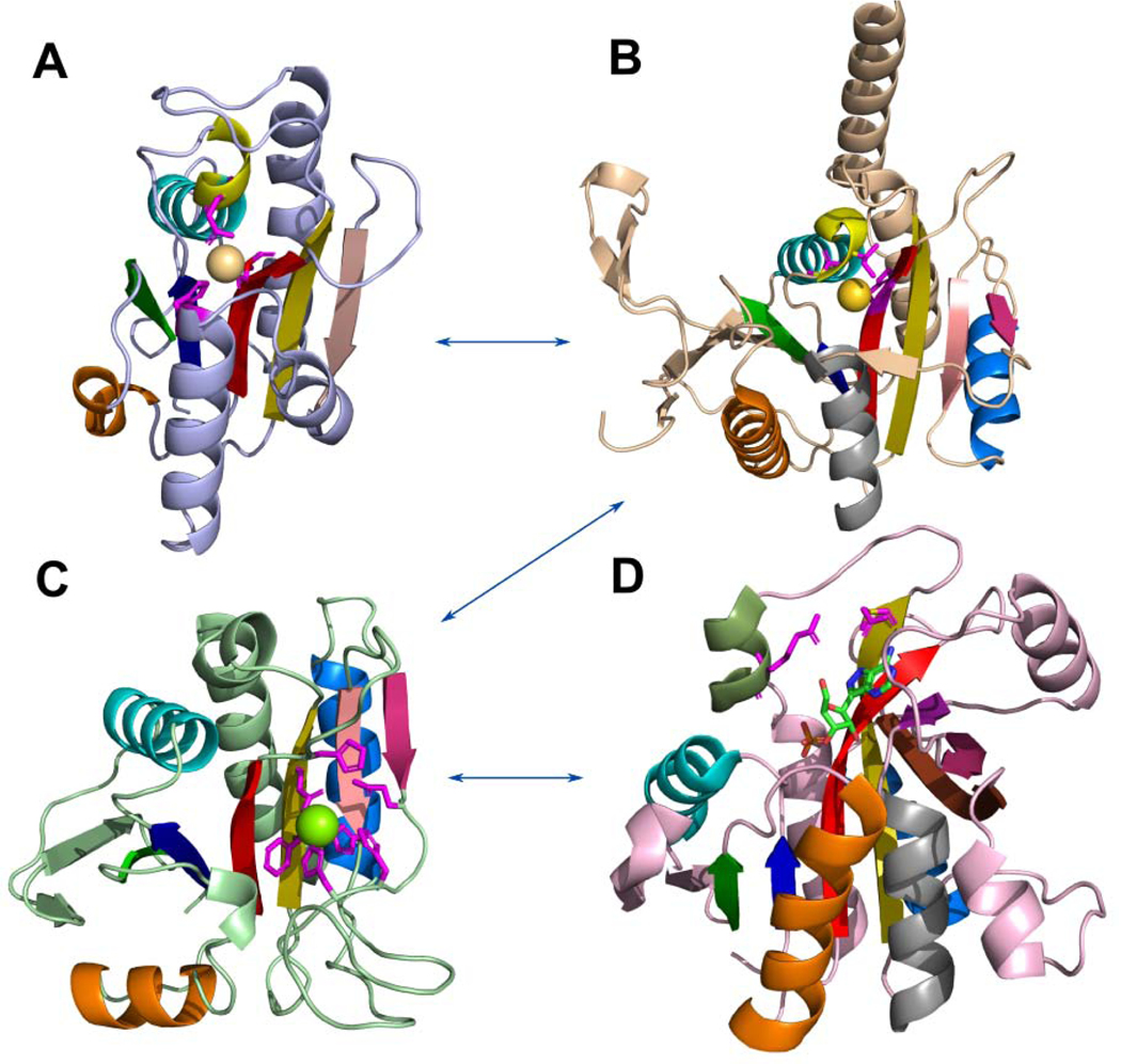
Fig 8. Probable evolutionary path between RLM-containing domains from Peptidyl-tRNA hydrolase-like homology group.
(A) Hydrogenase maturating endopeptidase from E.coli (PDB: 1cfz); (B) archaeal proteasome activator from Pyrococcus furiosus (PDB: 3vr0); (C) PH0006 protein from Pyrococcus horikoshii (PDB: 2gfq); (D) purine nucleoside phosphorylase from E.coli (PDB: 4ts3), formycin A is shown by sticks and colored by elements. RLM is shown in rainbow. 3(10)-helix is colored in yellow in A and B. Metal-binding residues are shown by sticks in A, B. Residues near metal-binding site are shown by sticks in C.

