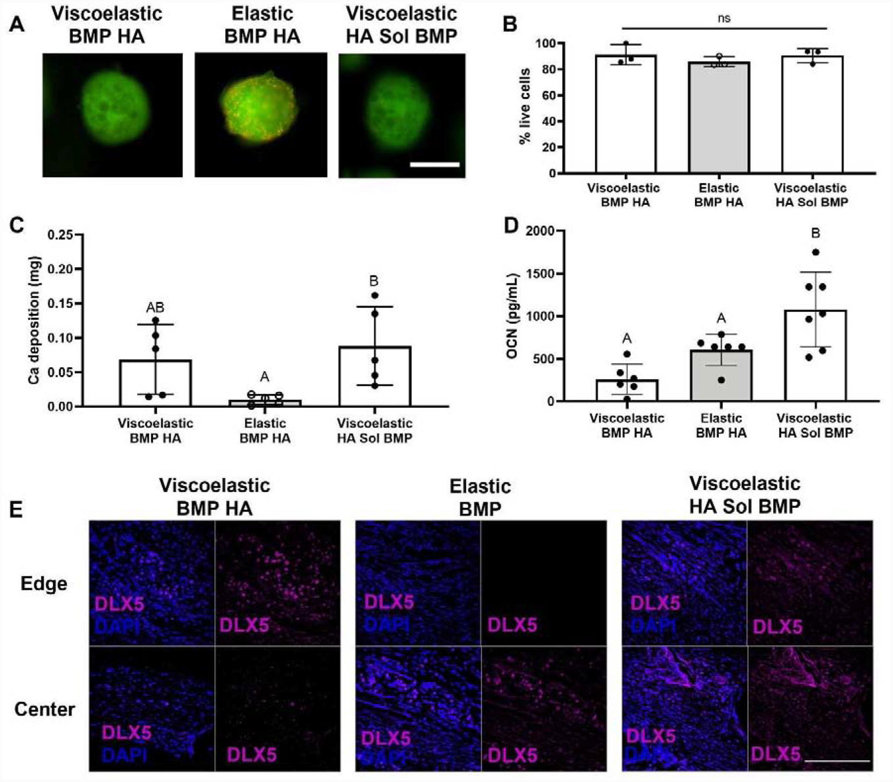
Figure 4. The viability and osteogenic potential of MSC spheroids is enhanced in viscoelastic alginate.

(A) Representative images of spheroids after 14 days in culture. Live cells are green and red cells are dead. Scale bar is 500 μm. (B) Quantification of live cells after 14 days in culture (n=3) (C) Calcium accumulation within viscoelastic and elastic alginate hydrogels over 14 days (n=5). (D) Cell-secreted osteocalcin after 14 days in culture (n=6–7). Significance is denoted by alphabetical letterings. Groups with statistically significant differences do not share the same letters. (E) Representative images of DLX5 staining (magenta) counterstained with DAPI (blue) from edge or center of the calvarial defect. Scale bar is 200 μm.
