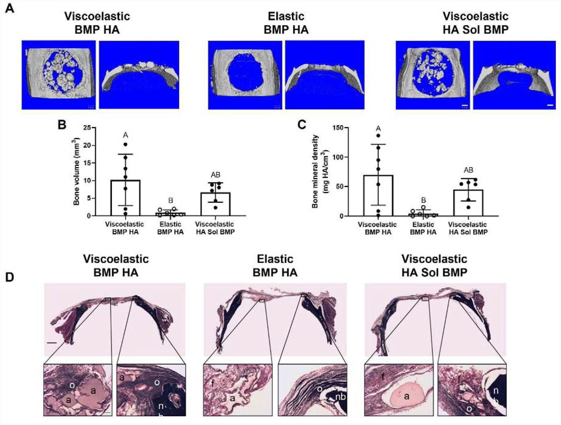
Figure 6. MSC spheroids promote bone tissue formation in vivo when entrapped in viscoelastic alginate.

(A) Representative images of microCT scans of explanted rat calvariae 12 weeks after hydrogel implantation. Scale bars are 1 mm. (B) Bone volume (n=6–7). (C) Bone mineral density (n=6–7). Significance is denoted by alphabetical letterings. Groups with statistically significant differences do not share the same letters. (D) Representative images of Masson’s Trichrome staining of calvarial defects from each group. Top images are large scans of an entire cross-section of the defect. Scale bar is 1 mm. The bottom insert images are 10X magnified regions of interest on either the center (left) or edge (right) of the defect. Residual alginate: a; fibrous tissue: f; native bone: nb; osteoid: o. Bottom scale bar is 200 μm.
