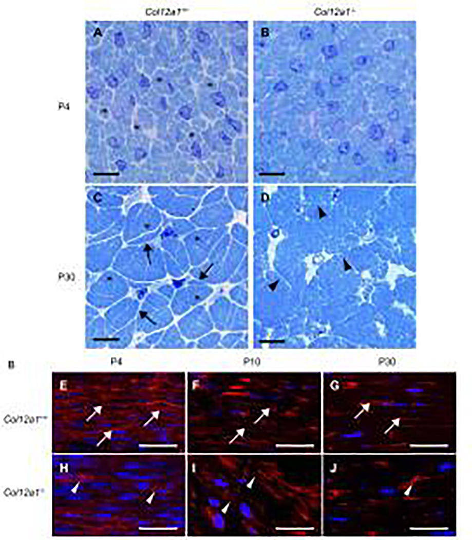
Fig 2. Tenocytes and fiber domain structure in cross and logitudinal sections.
(A-D) Cross sections of FDLs were stained with Toluidine blue and (E-J) longitudinal sections were stained with phalloidin and DAPI. At P4, Col12a1+/+ FDLs consisted of tenocytes and collagen fibers (asterisks). The domains, defined by the tenocytes containing fibers, are clearly defined (A), whereas no clear fiber domains are detected in Col12a1−/− FDLs (B). At P30, Col12a1+/+ tenocyte processes (arrows) interact with processes from neighboring cells and clearly define the fiber domains (asterisks) (C). In contrast, tenocyte processes (arrow heads) and fiber domains are disorganized and poorly defined in Col12a1−/− FDLs (D). Phalloidin staining of longitudinal sections demonstrates that tenocytes (arrows) are parallel and orientated along with the longitudinal axis at P4 (E). At P10 and P30, the tenocytes become attenuated along the tendon axis (F, G). In Col12a1−/− FDLs at P4, the actin cytoskeleton is less developed, and tenocytes are poorly organized (arrowheads) (H). Tenocyte structure (cytoskeleton) is disrupted, and the tenocytes are disorganized with the tendon axis hard to define at P10 (I) and P30 (J). Scale bars 50 μm (A-D) and 25 μm (E-J).

