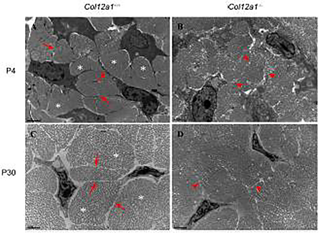
Fig 3. Altered tenocyte process formation and fibril spacing in the absence of collagen XII.
FDL cross sections from P4 and P30 Col12a1+/+ and Col12a1−/− mice were analyzed by transmission electron microscopy (TEM). At P4, Col12a1+/+ tenocytes extend their processes (arrows) and interact with adjacent cells. Fibers (asterisks) are surrounded by tenocyte processes (A). In contrast, tenocyte processes (arrowheads) are unclear and no obvious, well defined fibers are observed in Col12a1−/− FDLs (B). At P30, similar to P4, Col12a1+/+ tenocytes have fine processes (arrows) and clear fiber domains (asterisks) (C), whereas no clear tenocyte processes (arrowheads) and fiber domains are found in the Col12a1−/− FDL (D). Scale bars 2 μm.

