
Figure 2.
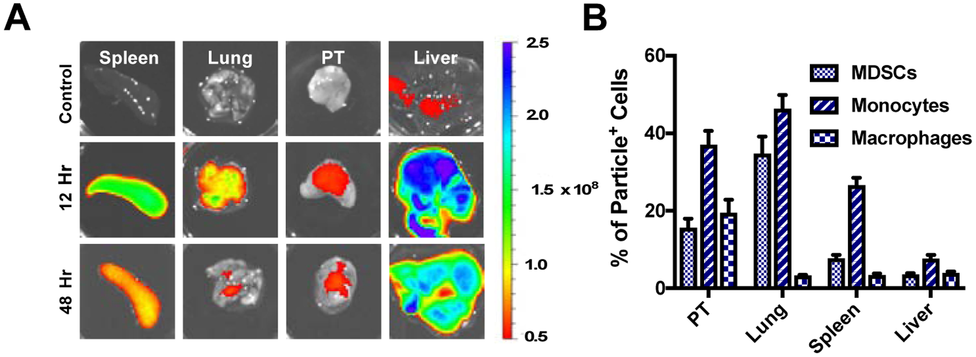
In vivo distribution of Cy5.5-NPs. (A) Ex vivo imaging following i.v. injection of 1 mg of Cy5.5-NPs shows they primarily accumulate in spleen and liver, and are detectable for more than 48 hours post injection. (B) Quantification of particle internalization by myeloid cell subsets within tissues as a percentage of total NP+ cells for a given tissue.