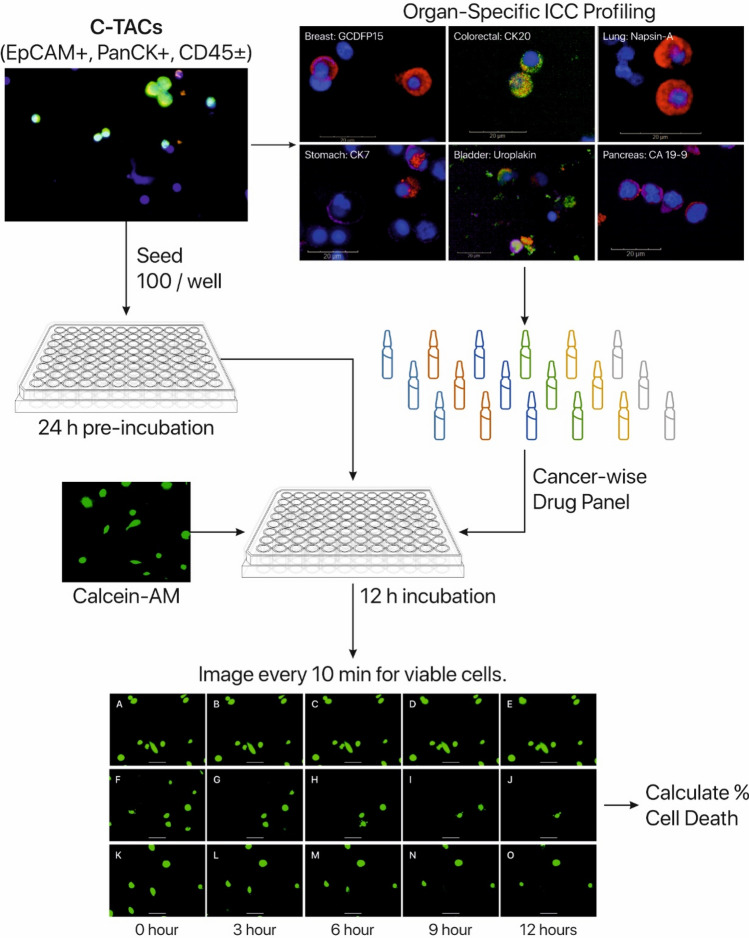
Fig. 2.
In vitro CRP workflow. C-TACs were ascertained by ICC profiling with OSS markers to identify cancer-specific drug panel. C-TACs were seeded into multi-well assay plates, pre-incubated and treated with appropriate CCA panel. C-TACs are stained with Calcein-AM to monitor viable cells during time-lapse fluorescent imaging where images were obtained every 10 min for 12 h. Proportion of surviving C-TACs were estimated to determine % cell death. Panels A-O show representative images of surviving C-TACs at various time points, when treated with different drugs with either low/no cytotoxicity (a–e), high cytotoxicity (f–j) and moderate cytotoxicity (k–o). Also see Supplementary video

