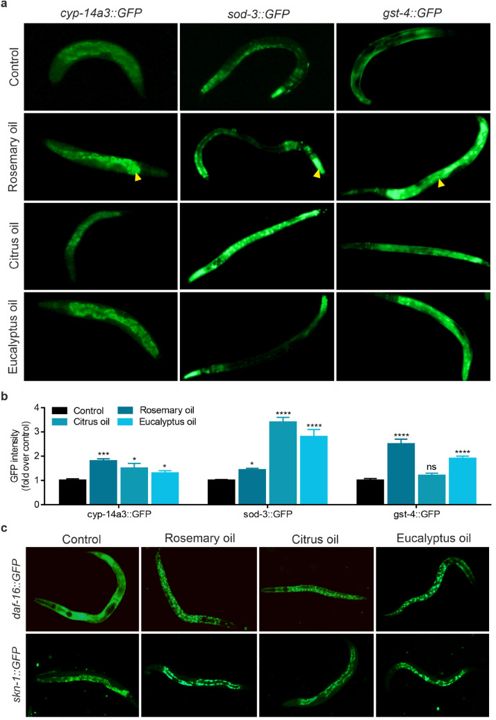
Fig. 7.
Expression and localization analysis of transgenic genes using GFP-reporter strains. Synchronized nematodes were treated with 0.25% [v/v] of EO or left untreated (control) for 72 h prior imaging. a Representative fluorescence microscopy images of cyp-14a3::GFP, sod-3::GFP and gst-4::GFP transgenic nematodes are shown. Arrow heads indicate characteristic expression pattern of the respective reporter gene. b Quantitation of indicated GFP-reporter gene intensity relative to control. Error bars are based on the SE of two independent experiments with at least 20 worms in each treatment. * p < 0.05, *** p < 0.001 and **** p < 0.0001 for comparison of EO treatment with control groups; ns, not significant. c Representative fluorescence microscopy images showing subcellular translocation behaviour of daf-16::GFP and skn-1::GFP upon three hours of EO (0.25% [v/v]) treatment

