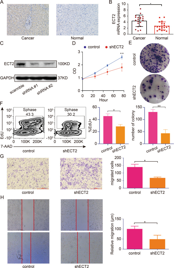
Fig. 3. Downregulation of ECT2 inhibits the proliferation and migration of HCC cells.
A Representative immunohistochemistry stain of ECT2 in HCC and para-cancerous normal tissues. B Quantification of PCR analysis of ECT2 expression in HCC and para-cancerous normal tissues. C Western blot analysis of ECT2 after transfection of ECT2-targeting shRNA. D ECT2 downregulation inhibits cell proliferation. Cancer cells were transfected with ECT2-targeting shRNA, and the absorption (A450 nm) was detected at 0, 24, 48, and 72 h. E Cancer cells transfected with ECT2-targeting shRNA and the normal control were assayed for clonogenicity in adherent cultures. F EdU incorporation assay was used to examine the proliferation of cancer cells after ECT2 knockdown. G Transwell assay was used for cancer cells transfected with ECT2-targeting shRNA (magnification 100×). H Wound-healing assay was used for cancer cells transfected with ECT2-targeting shRNA (magnification 50×, scale bar: 500 μm). Error bars represent means ± SD. *p < 0.05, **p < 0.01. NS means “not significant” by paired two-sided Student’s t-test.

