Figure 3.

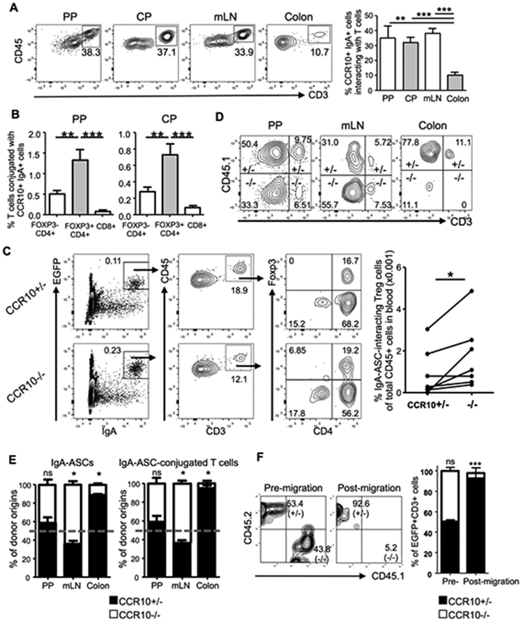
CCR10 mediates co-migration of IgA-ASCs and conjugating T cells into the colon. (A) Analysis of gated EGFP(CCR10)+CD45+intracellular IgA+ cells of the PP, CP, mLN and colons of CCR10+/EGFP mice to detect interacting CD3+ T cells. The bar graph on the right shows average percentages of CCR10(EGFP)+IgA+ cells that interact with T cells. 6 mice for PP and CP, 10 for mLN and 8 for colon were analyzed in at least 3 independent experiments. (B) Comparison of percentages of different subsets of T cells in the PP and CP of CCR10+/− mice that form conjugates with EGFP(CCR10)+ IgA-ASCs. 6 mice were analyzed. (C) Analysis of gated blood CD45+ cells of CCR10+/− and CCR10−/− mice for detection of interacting CCR10(EGFP)+intracellular IgA+ ASCs and T cells. Average percentages of Treg cells that conjugate with EGFP(CCR10)+ IgA+ cells of total CD45+ cells in the blood are presented in the bar graph on the right. One dot represents one mouse. Pairs of mice analyzed in parallel in same experiments are linked by lines. (D) Representative flow cytometric analysis of gated colonic EGFP+ intracellular IgA+ cells for their CCR10+/− (CD45.1+) versus CCR10−/− (CD45.1−) donor B cell origins and their interacting CD3+ T cells in the PP, mLN and colonic LP in μMT recipient mice two weeks after transfer. (E) Relative percentages of contribution of CCR10−/− (−/−) versus CCR10+/− (+/−) donor B cells to total donor-derived EGFP+ IgA-ASCs (left panel) and their interacting T cells (right panel) in PP, mLN and colon of μMT recipients two weeks after transfer. The relative percentages of contribution of CCR10+/− versus CCR10−/− donor B cells to IgA-ASCs were normalized on ratios of CCR10+/− versus CCR10−/− donor B cells in spleens of the recipients. The dashed line indicates the 50% of relative contribution from each donor origin if CCR10+/− and CCR10−/− donor cells contribute equally. Total 8 recipient mice were analyzed. (F) Flow cytometric analysis of 1:1 mixture of gated intestinal EGFP+CD3+ IgA-ASC/T cell conjugates of CCR10+/− and CCR10−/− mice (pre-migration) and cells recovered in bottom of a transwell after migration towards CCL28 in an in vitro migration assay (post-migration). Cells of 3 mice of each genotype were analyzed.
