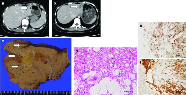
Fig. 2.

Images of case 2. a Contrast-enhanced CT image obtained in case 2 at the time of the first surgery (for the right mature cystic teratoma). The tumor measured 10 mm in diameter (arrow: the tumor). b Contrast-enhanced CT image obtained in case 2 at 2 years after the first surgery. The tumor measured 20 mm in diameter and possessed various components, including an adipose component (arrow: the tumor). c Resected specimen from case 2. The cut surface of the resected specimen contained a light brownish and yellow tumor, which measured 20 mm in diameter (arrow: the tumor). d HE staining of the tumor in a high-power field. The tumor consisted of a mixture of adipocytes, spindle-shaped cells, and epithelioid cells. Large atypical epithelioid cells with eosinophilic to clear cytoplasm were also identified. e HMB-45 staining of the tumor in a low-power field. The tumor was positive for HMB-45. f: α-SMA staining of the tumor in a low-power field. The tumor was positive for α-SMA
