Fig. 6.
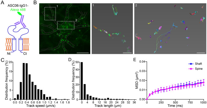
Lateral mobility of neuronal surface hASIC1a. A Diagram of direct labeling of surface hASIC1a by Alexa 488-conjugated ASC06-IgG1. B–D SPT experiments on neuronal surface hASIC1a. Asic1a−/− neurons transfected with hASIC1a were incubated with a low concentration of ASC06-IgG1-Alexa 488. SPT was performed for 1 min–2 min using TIRF microscope at 20 Hz. B Representative image of a neuron labeled by ASC06-IgG1-Alexa 488 under live conditions (left panel) and enlarged reconstructed trajectories of surface hASIC1a clusters in the boxed areas (middle and right panels). Scale bars: left panel, 10 μm; middle and right panels, 1 μm. C, D Quantification of SPT on distribution frequencies of track speeds (C) and track lengths (D) of surface hASIC1a particles (n = 681 particles counted). Track speed = track length/track time. E Mean square displacement (MSD) as a function of time for surface hASIC1a clusters on dendritic shafts and spines. Data points represent the mean ± SEM of 17 cells. Paired t test on area under the curve showed no significant difference between shafts and spines.

