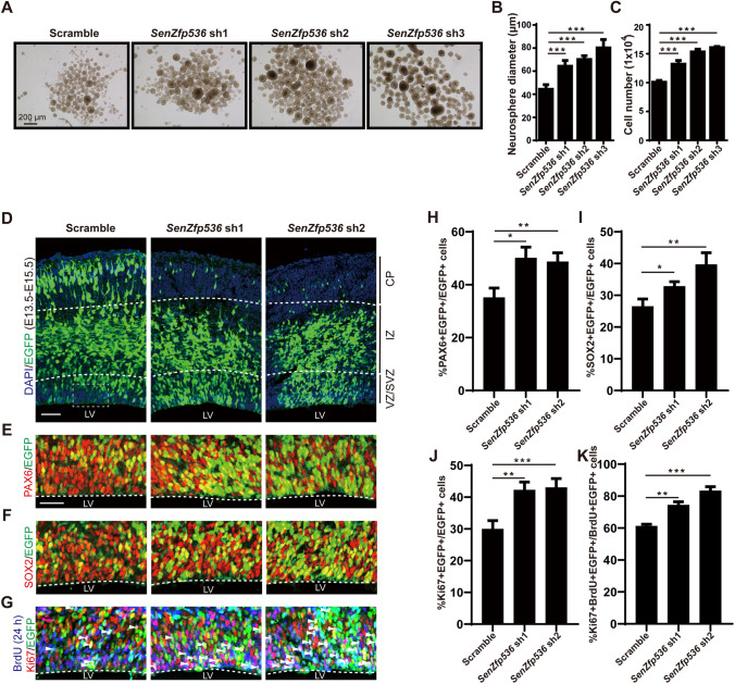
Fig. 6.
SenZfp536 regulates the proliferation of cortical NPCs. A Representative images showing E12.5 cortices cultured under sphere conditions and transduced with the indicated shRNAs for 7 days. B Bar plots of the average diameter of neurospheres as in A. F = 43.86, P < 0.0001 (one-way ANOVA followed by Dunnett’s multiple comparison test). C Bar plots of the cell numbers of neurospheres as in A. F = 112.4, P < 0.0001 (one-way ANOVA followed by Dunnett’s multiple comparison test). D–K E13.5 mouse cortices were electroporated with a mix of shRNA-expressing and EGFP-expressing vectors; embryos were sacrificed at E15.5 for immunofluorescent analyses. Spatial distributions of transduced cells (D). VZ/SVZ immunofluorescent images and quantification of PAX6+ (E, H), SOX2+ (F, I), and Ki67+ (G, J) transduced cells (EGFP+) are displayed. Co-immunostaining and quantification for 24-h BrdU (E14.5-E15.5), Ki67, and EGFP reveal that SenZfp536 knockdown leads to more cells staying in the cell cycle as measured by the percentages of BrdU+/Ki67+/EGFP+ among BrdU+/EGFP+ cells (G, K). For H, F = 30.30, P < 0.001; for (I, F = 34.05, P < 0.001; for J, F = 103.9, P < 0.0001; for K, F = 123.6, P < 0.0001 (one-way ANOVA followed by Dunnett’s multiple comparison test). Embryos in each experiment: scrambled, n = 4; SenZfp536 sh1, n = 4; SenZfp536 sh2, n = 3. Scale bars, 50 μm. *P < 0.05; **P <0.01; ***P < 0.001; ns, not significant. Results are presented as mean ± SEM.

