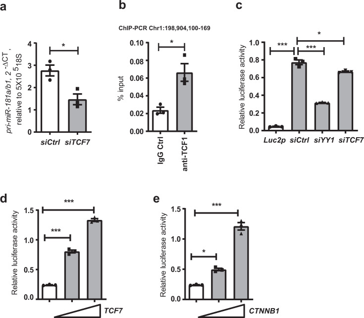
Fig. 1. Regulation of pri-miR-181a/b1 expression by TCF1.
a Naive CD4 T cells from young adults were transfected with TCF7 siRNA or control siRNA and assayed for pri-miR-181a/b1 expression after 48 h by q-PCR. Data are shown as mean±SEM (n = 3). b ChIP assay was performed on naïve CD4 T cells with anti-human TCF1 antibodies, and the sequence representing a previously identified pri-miR-181a/b1 enhancer region (chr1:198,904,100-169) was amplified. Precipitation with normal IgG was used as control. Data are shown as mean±SEM (n = 3). c Sequences corresponding to pri-miR-181a/b1 enhancer region (chr1:198,904,065-558) were cloned into a pGL4.27 [luc2P/minP/Hygro] plasmid. Dual-luciferase reporter assays were performed in HEK293T cells transfected with TCF7 siRNA, YY1 siRNA, or control siRNA. Data are shown as mean±SEM (n = 3). d, e Increasing amounts (0 ng, 30 ng or 100 ng) of TCF7 (d) and CTNNB1 (e) –containing plasmids were co-transfected with the pri-miR-181a/b1-enhancer-Luc2p reporter construct into HEK293T cells. Reporter activities after 48 h are shown as mean±SEM (n = 3). Comparisons were done by two-tailed paired t test in a, b; or, by one-way ANOVA with post-hoc Tukey test in c, d, and e. Significance levels are indicated as *P < 0.05, **P < 0.005, ***P < 0.0001.

